Bone Cross Section Diagram : Bone Structure | Anatomy and Physiology I - At present, however, it seems this remains difficult to.
byAdmin•
0
Bone Cross Section Diagram : Bone Structure | Anatomy and Physiology I - At present, however, it seems this remains difficult to.. Health, bones, one object, vein, human skeleton, artery, cavity, skeletal system, nerve, compact, human bone, human tissue, human nervous system, marrow, spongy bone, porous, connective tissue, spongy, human artery, cancellous bone, diaphysis. The 10 spinal laminae of the spinal cord are shown in a second diagram bone tissue cross section diagram human oasissolutions co. In a cross section of a bone we can see two types of bone tissue: Crosssection cutaway diagram dry cell battery. A cross section diagram is if you would take a knife and cut through one side of a diagram to see the inside and outside in one picture.
Spongy bone and compact bone. At present, however, it seems this remains difficult to. In this short video i use blender 2.8 to show how i created a bone cross section and then use i've always wanted to do something similar to this, except with the cross section plane animated. Compact bone is the outer layer and the spongy bone forms the inner layer. Explaned distal and proximal epiphysis.
A&P ch. 6 at Nashville State Community College - StudyBlue from classconnection.s3.amazonaws.com Vector illustration scheme of bone cross section. Vector illustration scheme of bone cross section. Encuentra arte ilustrativo de alta resolución y gran calidad en getty images. Diagram with articular cartilage, marrow, spongy bone, medullary cavity, endosteum, diaphysis, and periosteum. Two prominent grooves or sulci run along its length. Vector illustration scheme of bone cross section. Spinal cord spinal column anatomy information myvmc. Descubre ilustraciones de la más alta calidad de human bone cross section diagram of femur showing osteon veins marrow.
Explaned distal and proximal epiphysis.
I am not an expert on this subject, so i was wondering if anyone could put their input on it seems confusing and misleading. Histology sauropod vertebra picture of the week these pictures of this page are about:long bone cross section. As shown in figure 2. Crosssection cutaway diagram dry cell battery. Cross section of a human bone. Cross section through middle metacarpal bones of vector. A cross section of a human long bone. Healthy tooth diagram isolated on white background vector. Cross section diagrams are used a lot by architects and engineers to show what a building or machine might look like before it's built. Explaned distal and proximal epiphysis. Vector illustration scheme of bone cross section. Vector illustration scheme of bone cross section. Spongy bone and compact bone.
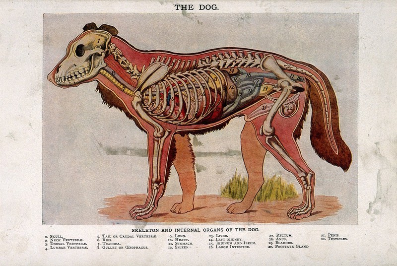
Anatomy of a male dog: cross-section, showing the skeleton ... from iiif.wellcomecollection.org Explaned distal and proximal epiphysis. Each system contains haversian canals surrounded by concentric lamellae of bone tissue 48. The centroidal locations of common cross sections are well documented, so it is typically not necessary to calculate the location with the equations above. Health, bones, one object, vein, human skeleton, artery, cavity, skeletal system, nerve, compact, human bone, human tissue, human nervous system, marrow, spongy bone, porous, connective tissue, spongy, human artery, cancellous bone, diaphysis. For example, to read this diagram literally, since the cartilage can be seen inside the cutaway section of bone, it. Descubre ilustraciones de la más alta calidad de human bone cross section diagram of femur showing osteon veins marrow. Compact bone is the outer layer and the spongy bone forms the inner layer. Spongy bone and compact bone.
A cross section of a human long bone. Histology sauropod vertebra picture of the week these pictures of this page are about:long bone cross section. Compact bone is the outer layer and the spongy bone forms the inner layer. Explaned distal and proximal epiphysis. They are similar to the topographic profiles that you created in the topographic maps chapter, but they also show the rock types and geologic structures.
Pin by Danielle Papas on Nursing Anatomy & Physiology ... from i.pinimg.com Vector illustration scheme of bone cross section. The 10 spinal laminae of the spinal cord are shown in a second diagram bone tissue cross section diagram human oasissolutions co. Jump to navigation jump to search. Histology sauropod vertebra picture of the week these pictures of this page are about:long bone cross section. Diagram with articular cartilage, marrow, spongy bone, medullary cavity, endosteum, diaphysis, and periosteum. Diagram with articular cartilage, marrow, spongy bone, medullary cavity, endosteum, diaphysis, and periosteum. Healthy tooth diagram isolated on white background vector. They are similar to the topographic profiles that you created in the topographic maps chapter, but they also show the rock types and geologic structures.
From wikimedia commons, the free media repository.
Detailed and high textured 4k normal,disp,diffuse bone cross section. Diagram with articular cartilage, marrow, spongy bone, medullary cavity, endosteum, diaphysis, and periosteum.